乳腺癌细针穿刺标本中端粒酶活性的临床意义
中华肿瘤杂志 2000年第2期第22卷 临床研究
作者:金顺钱 张伟 滕茂芳 张智慧 刘毅 李茉 曲平 王淑珍 金玉生 王洪平 潘秦镜 刘树范
单位:金顺钱(中国医学科学院中国协和医科大学肿瘤研究所肿瘤医院肿瘤生物学检测中心,北京,100021);张伟(中国医学科学院中国协和医科大学肿瘤研究所肿瘤医院肿瘤生物学检测中心,北京,100021);滕茂芳(中国医学科学院中国协和医科大学肿瘤研究所肿瘤医院肿瘤生物学检测中心,北京,100021);张智慧(中国医学科学院中国协和医科大学肿瘤研究所肿瘤医院肿瘤生物学检测中心,北京,100021);刘毅(中国医学科学院中国协和医科大学肿瘤研究所肿瘤医院肿瘤生物学检测中心,北京,100021)
关键词: 乳腺肿瘤/病理学;乳腺肿瘤/诊断;免疫组织化学;端粒酶;聚合酶链反应
【摘要】 目的 研究乳腺细针穿刺细胞标本中的端粒酶活性,探讨其在乳腺癌辅助诊断中的临床意义;研究端粒酶表达与PR、ER、PCNA、c-erbB2及p53之间的可能关系。方法 采用PCR-TRAP技术检测99例乳腺细针穿刺标本中的端粒酶活性。采用免疫组化技术检测35例乳腺癌组织标本中孕激素受体(PR)、雌激素受体(ER)、细胞增殖核抗原(PCNA)、癌基因c-erbB2及p53蛋白表达水平。结果 83例乳腺癌标本,在69例细胞学阳性标本中61例端粒酶阳性,7例细胞学可疑标本中5例端粒酶阳性,7例细胞学阴性标本中4例端粒酶阳性,端粒酶敏感性为84.3%(70/83),端粒酶活性检测和细胞学诊断总符合率为77.1%(64/83);12例乳腺良性病变标本中,4例端粒酶阳性;4例乳腺炎标本均为阴性。端粒酶表达与乳腺癌组织学类型、淋巴结转移、肿瘤大小、临床分期无相关性,与PR、ER状态、PCNA、c-erbB2表达亦无相关性,但与p53蛋白表达水平呈负相关。结论 乳腺细针穿刺标本端粒酶活性检测在乳腺癌临床诊断中有辅助诊断价值;其与p53蛋白表达水平之间的关系有待进一步阐明。
Clinical implications of telomerase activity in breast cancer fine-needle aspirates
JIN Shunqian, ZHANG Wei, TENG Maofang, et al.
(Cancer Institute (Hospital), Chinese Academy of Medical Sciences, Peking Union Medical College, Beijing 100021, China)
【Abstract】 Objective To study the telomerase activity in samples of breast fine-needle aspirates, its clinical implications, and possible association with expression of PR, ER, PCNA, c-erbB2 and p53.Methods Ninetynine fine needle aspirates from breast cancers in 83 patients, 12 from benign lesions and 4 from inflammation of the breast, were examined for telomerase activity by a PCR-based telomere repeat amplification protocol (PCR-TRAP). In 35 breast cancer tissue specimens, expression of PR, ER, PCNA, c-erbB2 and p53 were examined by immunohistochemical staining.Results Among 83 fine-needle aspirates from breast cancer, telomerase activity was detected in 61 of 69 cytology-possitive specimens and in 5 of 7 cytology-suspicious specimens and in 4 of 7 cytology-negative specimens, with an overall positive rate of 84.3% (70/83). The concordance rate of telomerase detection and cytologic examination was 77.1% (64/83). Of 12 fine-needle aspirates from patients with benign breast lesions, 4 were telomerase positive, but 4 aspirates from inflammed breast were all negative. Telomerase activity of breast cancer aspirates did not correlate with histologic type, lymph node status, tumor size and clinical stage, nor did it correlate with PR, ER status and PCNA and c-erbB2 expression. However, negative correlation was found between telomerase activity and accumulation of p53 proteins.Conclusion Detection of telomerase in breast fine-needle aspirates complementary to cytological examination is useful in the diagnosis of breast cancer. Its possible association with p53 accummulation needs further study.
【Subject words】 Breast neoplasms/pathology; Breast neoplasms/diagnosis; Immunohistochemistry; Telomerase; Polymerase chain reaction
端粒和端粒酶在肿瘤发生、诊断、治疗及预后判断等研究中均具有重要地位[1]。我们研究乳腺细针穿刺细胞标本中的端粒酶活性,旨在探讨端粒酶活性检测在乳腺癌生物学诊断中的临床意义。
材料与方法
1.标本来源:99例乳腺细针穿刺细胞标本,包括83例乳腺癌,12例乳腺良性病变(乳腺腺瘤7例,乳腺腺病2例,乳腺增生2例,导管内乳头状瘤1例)和4例乳腺炎标本,均取自我院1997年9月~1998年9月的就诊患者,全部病例均经我院病理证实。
2.细胞学诊断:由我院细胞学室按常规细胞学诊断方法做出。
3.免疫组织化学检测:采用常规免疫组化技术,分别对乳腺癌手术切除标本的孕激素受体(PR)、雌性激素受体(ER)、细胞增殖核抗原(PCNA)、癌基因c-erbB2及p53蛋白的表达情况进行检测。抗体购自Dako公司。结果判定标准:阳性细胞数≤5%者为-,6%~25%者为+,26%~50%之间者为++,51%~75%之间者为+++,>76%者为++++。
4.端粒酶提取:细针穿刺细胞标本加入100 μl预冷的端粒酶裂解液[10 mmol/L Tris-HCl (pH7.5),1 mmol/L MgCl2,1 mmol/L EGTA,5 mmol/L 二巯基乙醇,0.5% CHAPS,10%甘油],0℃裂解30 min,13 500×g 4℃离心30 min,吸取上清至一新预冷管中,定量后标本-80℃冻存。
5.端粒酶活性检测:参照TRAP方法[2]检测端粒酶活性。TS引物:5′-AATCCGTCGAGCAGAGTT-3′;CX引物:5′-CCCTTACCCTTACCCTTACCCTAA-3′。 反应条件为94℃ 40 s,50℃ 40 s, 72 ℃50 s,31个循环,最后72℃延长10 min。PCR产物经10%聚丙烯酰胺凝胶分离,-80℃放射自显影。阳性对照:宫颈癌HeLa细胞系蛋白提取物;阴性对照:无蛋白PCR反应液。显示3条以上梯形条带者判定为阳性。对阳性标本的第3条以上的阳性带用扫描仪进行光学扫描,并与HeLa细胞的检测结果作比较。比值<1/4者判定为低活性,>1/4者判定为高活性。
6.统计分析:以χ2检验进行统计分析。
结果
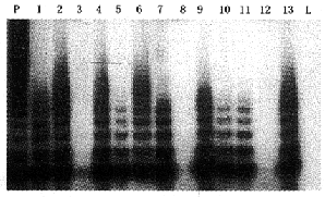
1.乳腺细针穿刺标本端粒酶诊断结果及与临床资料比较:4例乳腺炎标本端粒酶均阴性;12例乳腺良性病变标本4例端粒酶阳性,其中3例为低活性。83例乳腺癌标本中,70例端粒酶阳性,敏感性为84.3%。阳性标本中80.0%(56/70)表现为端粒酶高活性,20.0%(14/70)为低活性。乳癌组织端粒酶表达明显高于乳腺良性肿瘤及乳腺炎症组织(P<0.05)。端粒酶表达活性高低与肿瘤类型、淋巴结转移、肿瘤大小及临床分期等无关(表1,图1)。
表1 乳腺细针穿刺标本端粒酶活性检测结果
| 项目 |
病例数 |
端粒酶活性检测 |
| - |
低活性 |
高活性 |
阳性率(%) |
| 乳腺炎 |
4 |
4 |
0 |
0 |
0* |
| 乳腺良性病变 |
12 |
8 |
3 |
1 |
33.3* |
| 乳腺癌 |
83 |
13 |
14 |
56 |
84.3 |
| 侵袭性导管癌 |
75 |
11 |
12 |
52 |
85.3 |
| 髓样癌 |
2 |
0 |
1 |
1 |
- |
| 粘液癌 |
2 |
1 |
0 |
1 |
- |
| 乳头状瘤恶变 |
3 |
1 |
0 |
2 |
- |
| 大汗腺癌 |
1 |
0 |
1 |
0 |
- |
| 淋巴结转移 |
| 阴性 |
36 |
6 |
6 |
24 |
83.3 |
| 阳性 |
47 |
7 |
8 |
32 |
85.1 |
| 肿瘤大小 |
| <2 cm |
31 |
5 |
5 |
21 |
83.9 |
| ≥2 cm |
52 |
8 |
9 |
35 |
84.6 |
| 分期 |
| Ⅰ |
19 |
4 |
3 |
12 |
78.9 |
| Ⅱa |
28 |
4 |
5 |
19 |
85.7 |
| Ⅱb |
32 |
5 |
5 |
22 |
84.4 |
| Ⅲa |
4 |
0 |
1 |
3 |
100.0 |
*与乳腺癌组织相比,P<0.05

p:人子宫颈癌HeLa细胞系为阳性对照;L:端粒酶裂解液为阴性对照;1~13:乳腺癌细针穿刺细胞标本
图1 乳腺癌细针穿刺标本端粒酶活性检测结果
2.乳腺癌细针穿刺标本细胞学诊断和端粒酶诊断结果比较:在83例乳腺癌针吸标本中,细胞学诊断阳性的69例标本中有61例端粒酶阳性;细胞学诊断可疑的7例标本有5例阳性,细胞学诊断阴性的7例标本有4例阳性;端粒酶活性检测和细胞学诊断总符合率为77.1%(64/83),二者联合阳性率可升至93.9%(78/83)。
3.乳腺癌端粒酶活性表达与其它免疫组织化学标志的比较:35例乳腺癌手术标本做了PR、ER、PCNA、c-erbB2及p53蛋白的免疫组化检测。PR、ER、PCNA及c-erbB2与端粒酶活性表达无关(P>0.05);p53蛋白表达水平与端粒酶表达水平呈负相关(P<0.05,表2)。
表2 乳腺癌细针穿刺标本免疫组化与端粒酶活性
表达结果比较
| 免疫组化 |
例数 |
端粒酶活性表达 |
| - |
低活性 |
高活性 |
| PR |
| - |
4 |
0 |
1 |
3 |
| + |
10 |
2 |
1 |
7 |
| ++ |
8 |
1 |
1 |
6 |
| +++ |
13 |
3 |
2 |
8 |
| ER |
| - |
8 |
0 |
2 |
6 |
| + |
18 |
4 |
3 |
11 |
| ++ |
6 |
1 |
0 |
5 |
| +++ |
3 |
1 |
0 |
2 |
| PCNA |
| - |
1 |
0 |
0 |
1 |
| + |
7 |
2 |
1 |
4 |
| ++ |
11 |
1 |
0 |
10 |
| +++ |
16 |
3 |
4 |
9 |
| c-erbB2 |
| - |
12 |
4 |
1 |
7 |
| + |
5 |
0 |
0 |
5 |
| ++ |
6 |
0 |
2 |
4 |
| +++ |
12 |
2 |
2 |
8 |
| p53 |
| - |
20 |
0 |
1 |
19 |
| + |
8 |
2 |
2 |
4 |
| ++ |
3 |
3 |
0 |
0 |
| +++ |
4 |
1 |
2 |
1 |
| 合计 |
35 |
6 |
5 |
24 |
讨论
端粒酶能以自身RNA为模板,在染色体末端合成端粒序列,维持端粒长度。端粒酶激活与细胞永生化关系极为密切。研究结果表明,端粒酶是目前已知最具广谱和肿瘤特异性的肿瘤标志物之一[1]。目前,国外已将端粒酶活性检测应用于尿液、大肠冲洗液、口腔漱洗液及宫颈脱落细胞标本的肿瘤诊断研究中。
Hiyama等[3]检测了14例乳腺癌细针穿刺细胞标本,发现14例均为端粒酶阳性。Sugino等[4]报道15例乳腺癌针吸标本,有10例端粒酶阳性,而29例乳腺良性病变针吸标本仅有3例为弱阳性。最近Pearson等[5]报道,19例乳腺癌针吸标本有17(90%)例端粒酶阳性。我们对99例乳腺细针穿刺细胞标本检测端粒酶活性,发现4例乳腺炎标本端粒酶活性均为阴性,12例乳腺良性疾病标本有4例阳性,而在83例乳腺癌针吸标本中,端粒酶检出率为84.3%(70/83),与细胞学方法敏感性(83.1%,69/83)接近,且二者符合率为77.1(64/83)。在7例细胞学可疑标本和7例细胞学阴性标本中,有9例端粒酶阳性,说明这两种检测方法除具有良好的一致性外,尚有相当的互补性。
端粒酶激活是肿瘤发生的早期事件,因此,端粒酶活性检测有可能作为肿瘤早期诊断指标。本研究结果显示,约1/3乳腺良性病变组织中端粒酶活性增高,其是否在腺体早期癌变过程中起作用,尚待进一步研究。
p53是十分重要的抑癌基因,该基因的失活,在多种人类恶性肿瘤的发生发展过程中起重要作用,并同端粒酶一起参与细胞永生化过程。然而,p53基因失活是否与端粒酶活化过程有关,尚无文献报道。本研究结果表明,p53蛋白堆积与端粒酶表达之间存在负相关性(P<0.05),提示p53基因失活和端粒酶活化在乳腺癌发生过程中可能是相互独立的两条路线。有文献报道,某些p53突变子单独就能使乳腺上皮细胞永生化[6],在Rb/p16途径失活情况下,端粒酶活化也可使乳腺上皮永生化[7]。我们的研究结果也支持存在多种细胞永生化途径的结论。
本研究表明,乳腺细针穿刺标本端粒酶检测是敏感性高、特异性较好的肿瘤生物学检测技术,配合细胞学检查,可提高细胞学诊断的敏感性。
基金项目:中国医学科学院CMB院校长基金资助项目
作者单位:李茉(中国医学科学院中国协和医科大学肿瘤研究所肿瘤医院肿瘤生物学检测中心,北京,100021)
曲平(中国医学科学院中国协和医科大学肿瘤研究所肿瘤医院肿瘤生物学检测中心,北京,100021)
王淑珍(中国医学科学院中国协和医科大学肿瘤研究所肿瘤医院肿瘤生物学检测中心,北京,100021)
金玉生(中国医学科学院中国协和医科大学肿瘤研究所肿瘤医院肿瘤生物学检测中心,北京,100021)
王洪平(中国医学科学院中国协和医科大学肿瘤研究所肿瘤医院肿瘤生物学检测中心,北京,100021)
潘秦镜(细胞学室)
刘树范(细胞学室)
参考文献:
[1] Kim NW. Clinical implications of telomerase in cancer.Eur J Cancer,1997, 33:781-786.
[2] Kim NW, Piatyszek MA, Prowse KR, et al. Specific association of human telomerase activity with immortal cells and cancer. Science,1994, 266:2011-2015.
[3] Hiyama E, Gollahon L, Kataoka T, et al. Telomerase activity in human breast tumors. J Natl Cancer Inst,1996, 88:116-124.
[4] Sugino T, yoshida K, Bolodeoku J, et al. Telomerase activity in human breast cancer and benign breast lesions: diagnostic applications in clinical specimens, including fine needle aspirates. Int J Cancer,1996, 69: 301-306.
[5] Pearson AS, Gollagon LS, O′Neal NC, et al. Detection of telomerase activity in breast masses by fine-needle aspiration. Ann Surg Oncol, 1998,5:186-193.
[p6] Cao Y, Gao Q, Wazer DE, et al. Abrogation of wild-type p53-mediated transactivation is insufficient for mutant p53-induced immortalization of normal human mammary epithelial cells. Cancer Res,1997,57:5584-5589.
[7] Kiyono T, Foster SA, Koop JI, et al. Both Rb/p16INK4a inactivation and telomerase activity are requried to immortalize human epithelial cells. Nature,1998,396:84-88.
收稿日期:1999-02-05